Why Compare U-ULA Plates to GrowDex?
Limitations of U-ULA Plates
- Inefficient Spheroid Formation for Some Cell Types: Not all cell types efficiently form spheroids in U-ULA plates. Instead, these cells often remain in loose aggregates, leading to irregular morphology and inconsistent experimental results. This limitation is particularly problematic for researchers aiming to develop standardized 3D culture models.
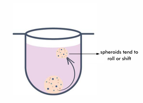
- Imaging Challenges Due to Spheroid Movement: In HCS and fluorescence imaging, spheroid stability is crucial for obtaining clear, high-resolution images. One major drawback of U-ULA plates is that spheroids tend to roll or shift, making it difficult to capture images from a consistent focal plane. This movement can introduce blur and inconsistencies in fluorescence signal detection, complicating multi-channel imaging and time-lapse studies. In contrast, hydrogel-based systems like GrowDex provide a stable environment that immobilizes spheroids, ensuring precise imaging over time.
- Lack of Cell-Matrix interactions: The extracellular matrix (ECM) is crucial for mimicking physiological cell-matrix interactions. Without ECM, spheroids rely solely on cell-cell adhesion, leading to weaker structural integrity, inaccurate tumor microenvironment modeling, and altered drug responses. ECM also supports cell migration and invasion, which U-ULA plates cannot replicate. This limitation affects tumor modeling and therapy testing, making ECM-based models more biologically relevant for cancer research and personalized medicine.
- Lower Cell Viability Over Extended Culture Periods: Short term cultures and treatments using U-ULA spheroids can work well, however for long-term cell culture experiments, spheroids generated in U-ULA plates may develop a necrotic core, and show lower viability compared to hydrogel-based scaffolds. This makes ULA plates less suitable for extended studies, particularly for research involving long-term drug exposure or tissue regeneration models.
While U-ULA plates are useful for high-throughput spheroid culture, their limitations in morphology control, reproducibility, imaging, and long-term viability make them less ideal for advanced 3D cell culture applications. Researchers seeking consistent, physiologically relevant models may benefit from hydrogel-based alternatives like GrowDex, which offer better spheroid formation, stability, and viability.
U-ULA Plates Vs GrowDex : A Comparison Study
GrowDex, a nanocellulose-based hydrogel, provides a 3D ECM like structure that enhances spheroid formation, maintains high viability, and improves imaging quality. Therefore, a comparison study was performed in house at UPM Biomedicals aiming to evaluate spheroid formation, viability, and reproducibility in U-bottom ULA plates versus GrowDex/GrowDex-T hydrogels across multiple cancer and normal cell lines.
Key Questions Answered in This Study:
- How does spheroid morphology compare between U-ULA plates and GrowDex?
- Do cells remain viable for longer in GrowDex?
- Which system provides more reproducible results across different experiments?
- How does imaging quality differ between the two systems?
Cell Line Performance: GrowDex vs. U-ULA
This study classified spheroids using the National Cancer Institute’s NCI60 categorization (Selby et al. 2017) based on the following categorisation:
- Category 1: Condensed spheroids
- Category 2: Spheroidal shapes
- Category 3: Aggregates
- Category 4: Loose aggregates
|
Cell Line
|
GrowDex/GrowDex - T
|
Category
|
U-ULA Plate
|
Category
|
|
PC-3 (Prostate Cancer)
|
Spheroidal structures
|
2
|
Loose aggregates
|
3
|
|
Panc-1 (Pancreatic Cancer)
|
Condensed spheroids
|
1
|
Spheroidal structures
|
2
|
|
Caki-1 (Renal Cancer)
|
Spheroidal structures, high viability
|
2
|
Spheroidal structures, lower viability
|
2
|
|
COLO205 (Colorectal Cancer)
|
Spheroidal structures, consistent morphology
|
2–3
|
Heterogeneous aggregates
|
2–4
|
|
A549 (Lung Cancer)
|
Condensed spheroids, uniform
|
1
|
Condensed spheroids, variable size
|
1
|
|
SK-MEL-5 (Melanoma)
|
Poor spheroid formation
|
|
Aggregates
|
3
|
|
IGROV-1 (Ovarian Cancer)
|
Aggregates
|
3
|
Loose aggregates
|
4
|
|
MCF7 (Breast Cancer)
|
Compact spheroids
|
1
|
Condensed spheroids/aggregates
|
1–3
|
|
mIMCD-3 (Mouse Kidney Cells)
|
Viable compact spheroids
|
1
|
Cells died by Day 4
|
|
Results from the Study
- Spheroid Morphology & Formation
The study demonstrated that GrowDex/GrowDex-T effectively supports compact spheroid formation, even in challenging cell types that failed to form spheroids in U-ULA plates. The natural nanocellulose matrix in GrowDex creates a more structured environment, leading to better cell-cell interactions and uniform spheroid growth. In contrast, U-ULA plates often resulted in loose aggregates, with spheroids displaying irregular morphology. GrowDex/T cultures also exhibited higher reproducibility, ensuring consistent spheroid formation across experiments, which is a crucial factor in drug discovery and cell-based screening.
- Viability & Longevity
Cell viability was significantly higher in GrowDex cultures, particularly in long-term experiments lasting 7–8 days. The hydrogel's extracellular matrix like properties provided structural and biochemical support that enhanced cell survival. In contrast, U-ULA cultures exhibited lower viability, with some cell lines showing early cell death. Notably, mIMCD-3 kidney cells died by Day 4 in U-ULA plates, while they remained viable and proliferative in GrowDex/GrowDex-T cultures. Additionally, some cell types, such as PANC-1 and PC3, showed better spheroid morphology and growth in GrowDex-T compared to standard GrowDex, highlighting the importance of hydrogel selection based on cell type.
- Imaging & High-Content Screening (HCS) Considerations
For HCS and fluorescence microscopy, GrowDex performed better than U-ULA plates. Using flat-bottom (F-bottom) plates with GrowDex resulted in better imaging clarity, as it provided a stable matrix that prevented spheroid movement. Conversely, U-bottom ULA plates had a small imaging window, making it difficult to capture high-resolution images. In live-cell imaging and fluorescence-based assays, spheroids in ULA plates tended to roll or shift, affecting the precision of time-lapse studies. By contrast, GrowDex immobilized spheroids, ensuring consistent positioning for repeated imaging and analysis, that is an essential feature for HCS workflows.
- Suitability for Different Cell Lines
The study revealed that GrowDex outperformed U-ULA plates in most tested cell lines, particularly in cancer and kidney-derived cells. While some cell lines, such as A549 lung cancer cells, showed similar morphology in both platforms, others, like MCF7 (breast cancer), PC3 (prostate cancer), and IGROV-1 (ovarian cancer), exhibited more compact and well-formed spheroids in GrowDex. Additionally, mIMCD-3 kidney cells struggled to survive in U-ULA plates, reinforcing that GrowDex provides superior support for long-term cultures.